
8 Nervous System
Learning Objectives
- Examine the anatomy of the nervous system
- Determine the main functions of the nervous system
- Differentiate the medical terms of the nervous system and common abbreviations
- Recognize the medical specialties associated with the nervous system
- Discover common diseases, disorders, and procedures related to the nervous system
Nervous System Word Parts
Click on prefixes, combining forms, and suffixes to reveal a list of word parts to memorize for the Nervous System.
Introduction to the Nervous System
The picture you have in your mind of the nervous system probably includes the brain, the nervous tissue contained within the cranium, and the spinal cord, the extension of nervous tissue within the vertebral column. That suggests it is made of two organs—and you may not even think of the spinal cord as an organ—but the nervous system is a very complex structure. Within the brain, many different and separate regions are responsible for many different and separate functions. It is as if the nervous system is composed of many organs that all look similar and can only be differentiated using tools such as the microscope or electrophysiology.
Watch this video:
Media 8.1 The Nervous System, Part 1: Crash Course A&P #8 [Online video]. Copyright 2015 by CrashCourse.
Practice Medical Terms Related to the Nervous System
Anatomy (Structures) of the Nervous System
The Central and Peripheral Nervous Systems
Nervous tissue, present in both the CNS and PNS, contains two basic types of cells: neurons and glial cells. Neurons are the primary type of cell that most anyone associates with the nervous system. They are responsible for the computation and communication that the nervous system provides. They are electrically active and release chemical signals to target cells. Glial cells, or glia, are known to play a supporting role for nervous tissue. Ongoing research pursues an expanded role that glial cells might play in signaling, but neurons are still considered the basis of this function. Neurons are important, but without glial support, they would not be able to perform their function. A glial cell is one of a variety of cells that provide a framework of tissue that supports the neurons and their activities. The neuron is the more functionally important of the two, in terms of the communicative function of the nervous system. To describe the functional divisions of the nervous system, it is important to understand the structure of a neuron.
Neurons are cells and therefore have a soma, or cell body, but they also have extensions of the cell; each extension is generally referred to as a process. There is one important process that every neuron has called an axon, which is the fiber that connects a neuron with its target. Another type of process that branches off from the soma is the dendrite. Dendrites are responsible for receiving most of the input from other neurons.
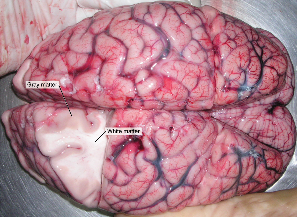
Looking at nervous tissue, some regions predominantly contain cell bodies and regions that are largely composed of just axons. These two regions within nervous system structures are often referred to as gray matter (the regions with many cell bodies and dendrites) or white matter (the regions with many axons). Figure 8.2 demonstrates the appearance of these regions in the brain and spinal cord. The colors ascribed to these regions are what would be seen in “fresh,” or unstained, nervous tissue. Gray matter is not necessarily gray. It can be pinkish because of blood content, or even slightly tan, depending on how long the tissue has been preserved. White matter is white because axons are insulated by a lipid-rich substance called myelin. Lipids can appear as white (“fatty”) material, much like the fat on a raw piece of chicken or beef. Gray matter may have that color ascribed to it because next to the white matter, it is just darker—hence, gray.
The distinction between gray matter and white matter is most often applied to central nervous tissue, which has large regions that can be seen with the unaided eye. When looking at peripheral structures, often a microscope is used and the tissue is stained with artificial colors. That is not to say that central nervous tissue cannot be stained and viewed under a microscope, but unstained tissue is most likely from the CNS—for example, a frontal section of the brain or cross-section of the spinal cord.
Did you know?
The brain has over 100 billion neurons.
The Adult Brain
The Cerebrum
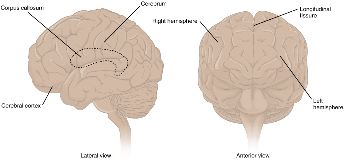
The iconic gray mantle of the human brain, which appears to make up most of the mass of the brain, is the cerebrum (see Figure 8.3). The wrinkled portion is the cerebral cortex, and the rest of the structure is beneath that outer covering. There is a large separation between the two sides of the cerebrum called the longitudinal fissure. It separates the cerebrum into two distinct halves, a right and left cerebral hemisphere. Deep within the cerebrum, the white matter of the corpus callosum provides the major pathway for communication between the two hemispheres of the cerebral cortex.
Many of the higher neurological functions, such as memory, emotion, and consciousness, are the result of cerebral function. The complexity of the cerebrum is different across vertebrate species. The cerebrum of the most primitive vertebrates is not much more than the connection for the sense of smell. In mammals, the cerebrum comprises the outer gray matter that is the cortex (from the Latin word meaning “bark of a tree”) and several deep nuclei that belong to three important functional groups. The basal nuclei are responsible for cognitive processing, the most important function being that associated with planning movements. The basal forebrain contains nuclei that are important in learning and memory. The limbic cortex is the region of the cerebral cortex that is part of the limbic system, a collection of structures involved in emotion, memory, and behavior.
Did you know?
The brain is about 75% water and is the fattest organ in the body.
Cerebral Cortex
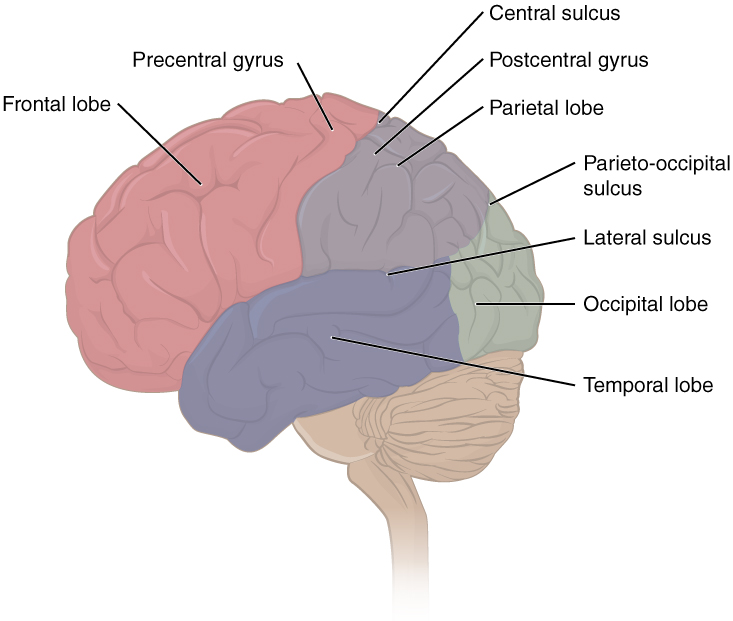
The cerebrum is covered by a continuous layer of gray matter that wraps around either side of the forebrain—the cerebral cortex. This thin, extensive region of wrinkled gray matter is responsible for the higher functions of the nervous system. A gyrus (plural = gyri) is the ridge of one of those wrinkles, and a sulcus (plural = sulci) is the groove between two gyri. The pattern of these folds of tissue indicates specific regions of the cerebral cortex.
The head is limited by the size of the birth canal, and the brain must fit inside the cranial cavity of the skull. Extensive folding in the cerebral cortex enables more gray matter to fit into this limited space. If the gray matter of the cortex were peeled off of the cerebrum and laid out flat, its surface area would be roughly equal to one square meter.
The folding of the cortex maximizes the amount of gray matter in the cranial cavity. During embryonic development, as the telencephalon expands within the skull, the brain goes through a regular course of growth that results in everyone’s brain having a similar pattern of folds. The surface of the brain can be mapped based on the locations of large gyri and sulci. Using these landmarks, the cortex can be separated into four major regions, or lobes (see Figure 8.4). The lateral sulcus that separates the temporal lobe from the other regions is one such landmark. Superior to the lateral sulcus is the parietal lobe and frontal lobe, which are separated from each other by the central sulcus. The posterior region of the cortex is the occipital lobe, which has no obvious anatomical border between it and the parietal or temporal lobes on the lateral surface of the brain. From the medial surface, an obvious landmark separating the parietal and occipital lobes is called the parieto-occipital sulcus. The fact that there is no obvious anatomical border between these lobes is consistent with the functions of these regions being interrelated.
Concept Check
- Identify the two major divisions of the nervous system.
- Describe the cerebral cortex.
- What are the halves of the cerebrum known as?
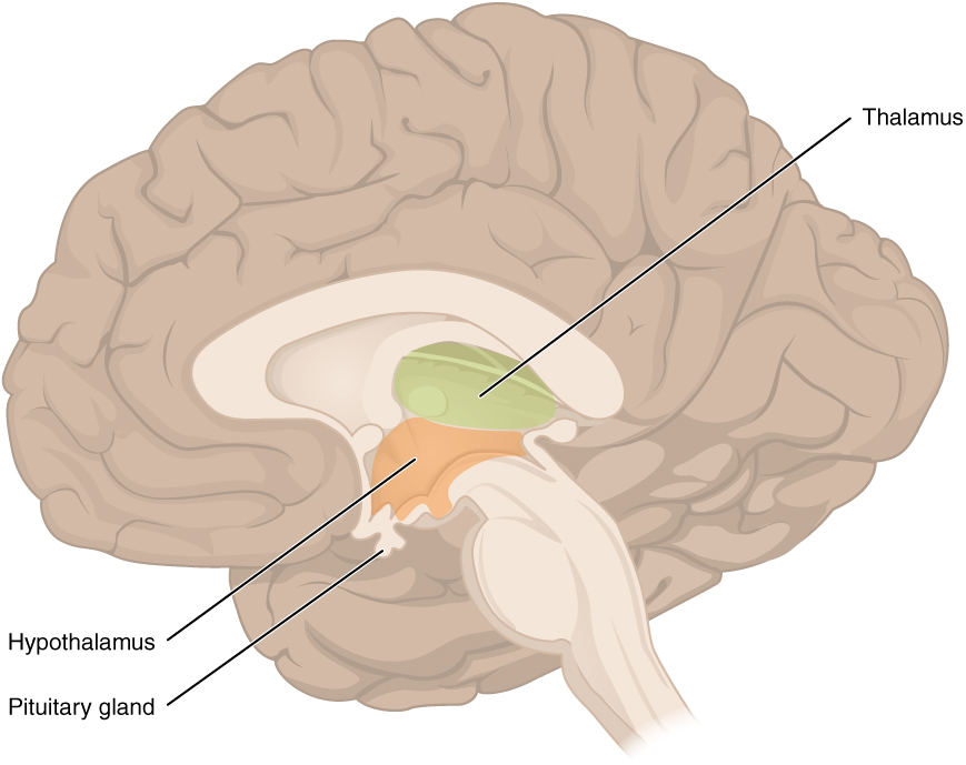
The Diencephalon
The diencephalon is deep beneath the cerebrum and constitutes the walls of the third ventricle. The diencephalon can be described as any region of the brain with “thalamus” in its name. The two major regions of the diencephalon are the thalamus itself and the hypothalamus (see Figure 8.5). There are other structures, such as the epithalamus, which contains the pineal gland, or the subthalamus, which includes the subthalamic nucleus that is part of the basal nuclei.
Thalamus
The thalamus is a collection of nuclei that relay information between the cerebral cortex and the periphery, spinal cord, or brainstem. All sensory information, except for the sense of smell, passes through the thalamus before processing by the cortex. For example, the portion of the thalamus that receives visual information will influence what visual stimuli are important, or what receives attention.
The cerebrum also sends information down to the thalamus, which usually communicates motor commands. This involves interactions with the cerebellum and other nuclei in the brainstem. The cerebrum interacts with the basal nuclei, which involves connections with the thalamus. The primary output of the basal nuclei is to the thalamus, which relays that output to the cerebral cortex. The cortex also sends information to the thalamus that will then influence the effects of the basal nuclei.
Hypothalamus
Inferior and slightly anterior to the thalamus is the hypothalamus, the other major region of the diencephalon. The hypothalamus is a collection of nuclei that are largely involved in regulating homeostasis. The hypothalamus is the executive region in charge of the autonomic nervous system and the endocrine system through its regulation of the anterior pituitary gland. Other parts of the hypothalamus are involved in memory and emotion as part of the limbic system.
Brain Stem
The midbrain and hindbrain (composed of the pons and the medulla) are collectively referred to as the brain stem (see Figure 8.6). The structure emerges from the ventral surface of the forebrain as a tapering cone that connects the brain to the spinal cord. Attached to the brain stem but considered a separate region of the adult brain is the cerebellum. The midbrain coordinates sensory representations of the visual, auditory, and somatosensory perceptual spaces. The pons is the main connection with the cerebellum. The pons and the medulla regulate several crucial functions, including the cardiovascular and respiratory systems and rates.
The cranial nerves connect through the brain stem and provide the brain with the sensory input and motor output associated with the head and neck, including most of the special senses. The major ascending and descending pathways between the spinal cord and brain, specifically the cerebrum, pass through the brain stem.
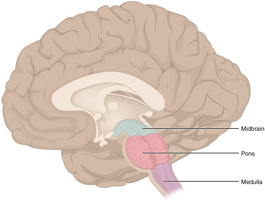
Midbrain
One of the original regions of the embryonic brain, the midbrain is a small region between the thalamus and pons. It is separated into the tectum and tegmentum, from the Latin words for roof and floor, respectively. The cerebral aqueduct passes through the center of the midbrain, such that these regions are the roof and floor of that canal.
Pons
The word pons comes from the Latin word for bridge. It is visible on the anterior surface of the brain stem as the thick bundle of white matter attached to the cerebellum. The pons is the main connection between the cerebellum and the brain stem. The bridge-like white matter is only the anterior surface of the pons; the gray matter beneath that is a continuation of the tegmentum from the midbrain. Gray matter in the tegmentum region of the pons contains neurons receiving descending input from the forebrain that is sent to the cerebellum.
Medulla
The medulla is the region known as the myelencephalon in the embryonic brain. The initial portion of the name, “myel,” refers to the significant white matter found in this region—especially on its exterior, which is continuous with the white matter of the spinal cord. The tegmentum of the midbrain and pons continues into the medulla because this gray matter is responsible for processing cranial nerve information. A diffuse region of gray matter throughout the brain stem, known as the reticular formation, is related to sleep and wakefulness, such as general brain activity and attention.
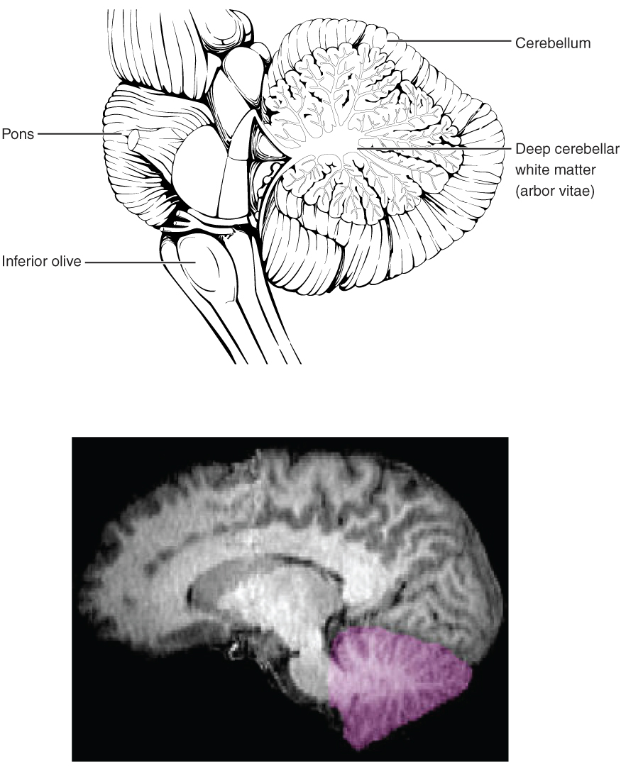
The Cerebellum
The cerebellum, as the name suggests, is the “little brain.” It is covered in gyri and sulci like the cerebrum and looks like a miniature version of that part of the brain (see Figure 8.7). The cerebellum is largely responsible for comparing information from the cerebrum with sensory feedback from the periphery through the spinal cord. It accounts for approximately 10% of the mass of the brain.
Concept Check
- What is the primary processing purpose of the medulla?
- Identify the structure in the brain responsible for sensory feedback through the spinal cord. Suggest what may happen if this function fails.
The Spinal Cord
The description of the CNS is concentrated on the structures of the brain, but the spinal cord is another major organ of the system. Whereas the brain develops out of expansions of the neural tube into primary and then secondary vesicles, the spinal cord maintains the tube structure and is only specialized into certain regions. As the spinal cord continues to develop in the newborn, anatomical features mark its surface. The anterior midline is marked by the anterior median fissure, and the posterior midline is marked by the posterior median sulcus. Axons enter the posterior side through the dorsal (posterior) nerve root, which marks the posterolateral sulcus on either side. The axons emerging from the anterior side do so through the ventral (anterior) nerve root. Note that it is common to see the terms dorsal (dorsal = “back”) and ventral (ventral = “belly”) used interchangeably with posterior and anterior, particularly in reference to nerves and the structures of the spinal cord. You should learn to be comfortable with both.
On the whole, the posterior regions are responsible for sensory functions and the anterior regions are associated with motor functions. This comes from the initial development of the spinal cord, which is divided into the basal plate and the alar plate. The basal plate is closest to the ventral midline of the neural tube, which will become the anterior face of the spinal cord and gives rise to motor neurons. The alar plate is on the dorsal side of the neural tube and gives rise to neurons that will receive sensory input from the periphery.
The length of the spinal cord is divided into regions that correspond to the regions of the vertebral column. The name of a spinal cord region corresponds to the level at which spinal nerves pass through the intervertebral foramina. Immediately adjacent to the brain stem are the following divisions of the spinal cord:
- cervical region
- thoracic region
- lumbar region
- sacral region.
The spinal cord is not the full length of the vertebral column because the spinal cord does not grow significantly longer after the first or second year, but the skeleton continues to grow. The nerves that emerge from the spinal cord pass through the intervertebral foramina at the respective levels. As the vertebral column grows, these nerves grow with it and result in a long bundle of nerves that resembles a horse’s tail and is named the cauda equina. The sacral spinal cord is at the level of the upper lumbar vertebral bones. The spinal nerves extend from their various levels to the proper level of the vertebral column.
Did you know?
The bundle of nerve fibers making up the spinal cord is no thicker than the human thumb.
Neurons
Neurons are the cells considered to be the basis of nervous tissue. They are responsible for the electrical signals that communicate information about sensations, and that produce movements in response to those stimuli, along with inducing thought processes within the brain. An important part of the function of neurons is in their structure or shape. The three-dimensional shape of these cells makes the immense number of connections within the nervous system possible.
Parts of a Neuron
As you learned in the first section, the main part of a neuron is the cell body, which is also known as the soma (soma = “body”). The cell body contains the nucleus and most of the major organelles. What makes neurons special is that they have many extensions of their cell membranes, which are generally referred to as processes. Neurons are usually described as having one, and only one, axon—a fiber that emerges from the cell body and projects to target cells. That single axon can branch repeatedly to communicate with many target cells. It is the axon that propagates the nerve impulse, which is communicated to one or more cells. The other processes of the neuron are dendrites, which receive information from other neurons at specialized areas of contact called synapses. The dendrites are usually highly branched processes, providing locations for other neurons to communicate with the cell body. Information flows through a neuron from the dendrites, across the cell body, and down the axon. This gives the neuron a polarity—meaning that information flows in this one direction. Figure 8.8 shows the relationship of these parts to one another.
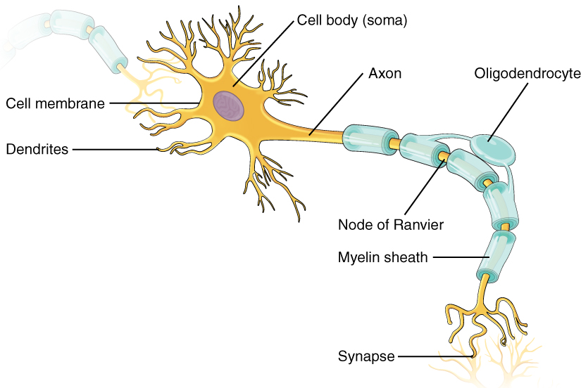
Where the axon emerges from the cell body, there is a special region referred to as the axon hillock. This is a tapering of the cell body toward the axon fiber. Within the axon hillock, the cytoplasm changes to a solution of limited components called axoplasm. Because the axon hillock represents the beginning of the axon, it is also referred to as the initial segment.
Many axons are wrapped by an insulating substance called myelin, which is made from glial cells. Myelin acts as insulation much like the plastic or rubber that is used to insulate electrical wires. A key difference between myelin and the insulation on a wire is that there are gaps in the myelin covering of an axon. Each gap is called a node of Ranvier and is important to the way that electrical signals travel down the axon. The length of the axon between each gap, which is wrapped in myelin, is referred to as an axon segment. At the end of the axon is the axon terminal, where there are usually several branches extending toward the target cell, each of which ends in an enlargement called a synaptic end bulb. These bulbs are what make the connection with the target cell at the synapse.
Types of Neurons
There are many neurons in the nervous system—a number in the trillions. And there are many different types of neurons. They can be classified by many different criteria. The first way to classify them is by the number of processes attached to the cell body. Using the standard model of neurons, one of these processes is the axon, and the rest are dendrites. Because information flows through the neuron from dendrites or cell bodies toward the axon, these names are based on the neuron’s polarity (see Figure 8.9).
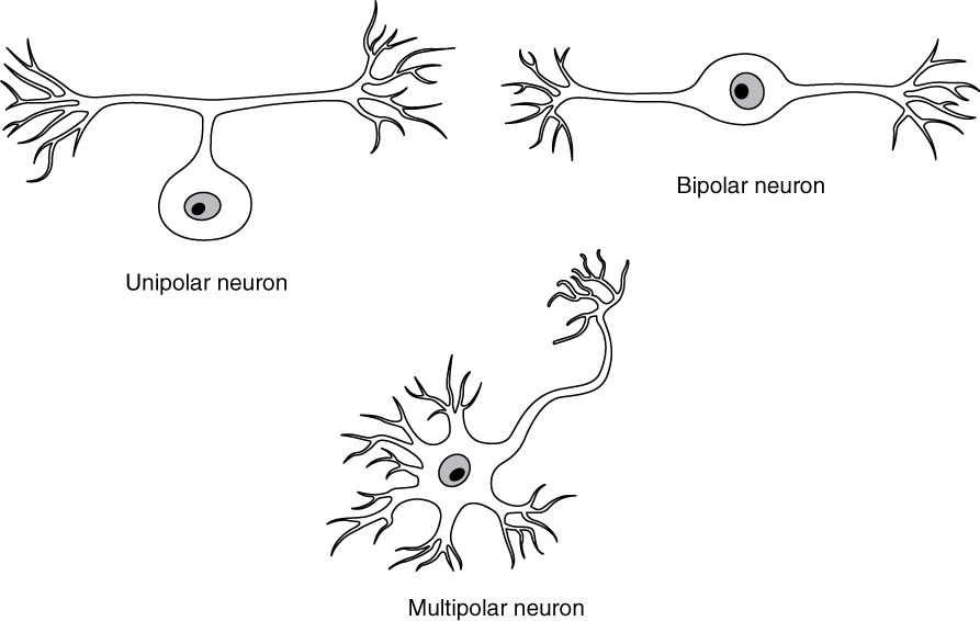
Unipolar cells have only one process emerging from the cell. True unipolar cells are only found in invertebrate animals, so the unipolar cells in humans are more appropriately called “pseudo-unipolar” cells. Invertebrate unipolar cells do not have dendrites.
Bipolar cells have two processes, which extend from each end of the cell body, opposite to each other. One is the axon and one the dendrite. Bipolar cells are not very common. They are found mainly in the olfactory epithelium (where smell stimuli are sensed), and as part of the retina.
Multipolar neurons are all of the neurons that are not unipolar or bipolar. They have one axon and two or more dendrites (usually many more). With the exception of the unipolar sensory ganglion cells, and the two specific bipolar cells mentioned above, all other neurons are multipolar.
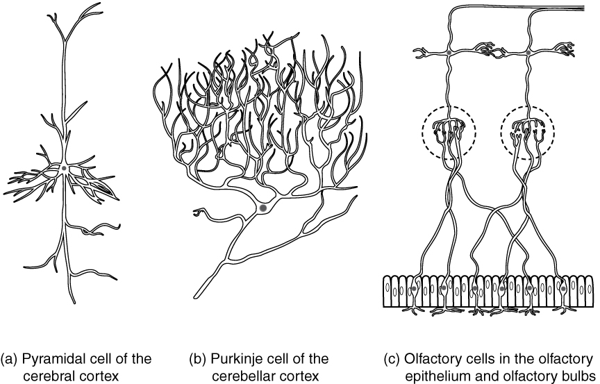
Neurons can also be classified on the basis of where they are found, who found them, what they do, or even what chemicals they use to communicate with each other. Some neurons referred to in this section on the nervous system are named on the basis of those sorts of classifications (see Figure 8.10). For example, a multipolar neuron that has a very important role to play in a part of the brain called the cerebellum is known as a Purkinje (commonly pronounced per-KIN-gee) cell. It is named after the anatomist who discovered it (Jan Evangilista Purkinje, 1787–1869).
Glial Cells
Glial cells, or neuroglia or simply glia, are the other type of cell found in nervous tissue. They are considered to be supporting cells, and many functions are directed at helping neurons complete their function for communication. The name glia comes from the Greek word that means “glue,” and was coined by the German pathologist Rudolph Virchow, who wrote in 1856, “This connective substance, which is in the brain, the spinal cord, and the special sense nerves, is a kind of glue (neuroglia) in which the nervous elements are planted.” Today, research into nervous tissue has shown that there are many deeper roles that these cells play, and research may find much more about them in the future.
There are six types of glial cells. Four of them are found in the CNS and two are found in the PNS. Table 8.1 outlines some common characteristics and functions.
| CNS GLIA | PNS GLIA | BASIC FUNCTION |
|---|---|---|
| Astrocyte | Satellite cell | Support |
| Oligodendrocyte | Schwann cell | Insulation, myelination |
| Microglia | – | Immune surveillance and phagocytosis |
| Ependymal cell | – | Creating CSF |
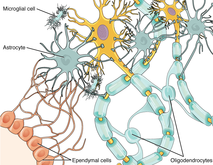
Glial Cells of the CNS
One cell providing support to neurons of the CNS is the astrocyte, so named because it appears to be star-shaped under the microscope (astro- = “star”). Astrocytes have many processes extending from their main cell body (not axons or dendrites like neurons, just cell extensions). Those processes extend to interact with neurons, blood vessels, or the connective tissue covering the CNS that is called the pia mater (see Figure 8.11). Generally, they are supporting cells for the neurons in the central nervous system. Some ways in which they support neurons in the central nervous system are by maintaining the concentration of chemicals in the extracellular space, removing excess signaling molecules, reacting to tissue damage, and contributing to the blood-brain barrier (BBB). The blood-brain barrier is a physiological barrier that keeps many substances that circulate in the rest of the body from getting into the central nervous system, restricting what can cross from circulating blood into the CNS. Nutrient molecules, such as glucose or amino acids, can pass through the BBB, but other molecules cannot. This actually causes problems with drug delivery to the CNS. Pharmaceutical companies are challenged to design drugs that can cross the BBB as well as have an effect on the nervous system.
Like a few other parts of the body, the brain has a privileged blood supply. Very little can pass through by diffusion. Most substances that cross the wall of a blood vessel into the CNS must do so through an active transport process. Because of this, only specific types of molecules can enter the CNS. Glucose—the primary energy source—is allowed, as are amino acids. Water and some other small particles, like gases and ions, can enter, but most everything else cannot, including white blood cells, which are one of the body’s main lines of defense. While this barrier protects the CNS from exposure to toxic or pathogenic substances, it also keeps out the cells that could protect the brain and spinal cord from disease and damage. The BBB also makes it harder for pharmaceuticals to be developed that can affect the nervous system. Aside from finding efficacious substances, the means of delivery is also crucial.
Oligodendrocyte, sometimes called just “oligo,” is the glial cell type that insulates axons in the CNS. The name means “cell of a few branches” (oligo- = “few”; dendro- = “branches”; -cyte = “cell”). There are a few processes that extend from the cell body. Each one reaches out and surrounds an axon to insulate it in myelin.
Microglia are, as the name implies, smaller than most of the other glial cells. Ongoing research into these cells, although not entirely conclusive, suggests that they may originate as white blood cells, called macrophages, that become part of the CNS during early development. While their origin is not conclusively determined, their function is related to what macrophages do in the rest of the body. When macrophages encounter diseased or damaged cells in the rest of the body, they ingest and digest those cells or the pathogens that cause disease. Microglia are the cells in the CNS that can do this in normal, healthy tissue, and they are therefore also referred to as CNS-resident macrophages.
The ependymal cell is a glial cell that filters blood to make cerebrospinal fluid (CSF), the fluid that circulates through the CNS. Because of the privileged blood supply inherent in the BBB, the extracellular space in nervous tissue does not easily exchange components with the blood. Ependymal cells line each ventricle, one of four central cavities that are remnants of the hollow center of the neural tube formed during the embryonic development of the brain. They also have cilia on their apical surface to help move the CSF through the ventricular space. The relationship of these glial cells to the structure of the CNS is seen in Figure 8.11.
Glial Cells of the PNS
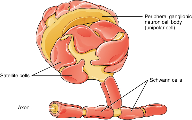
One of the two types of glial cells found in the PNS is the satellite cell. Satellite cells are found in sensory and autonomic ganglia, where they surround the cell bodies of neurons. This accounts for the name, based on their appearance under the microscope. They provide support, performing similar functions in the periphery as astrocytes do in the CNS—except, of course, for establishing the BBB.
The second type of glial cell is the Schwann cell, which insulates axons with myelin in the periphery. Schwann cells are different from oligodendrocytes in that a Schwann cell wraps around a portion of only one axon segment and no others. Oligodendrocytes have processes that reach out to multiple axon segments, whereas the entire Schwann cell surrounds just one axon segment. The nucleus and cytoplasm of the Schwann cell are on the edge of the myelin sheath. The relationship of these two types of glial cells to ganglia and nerves in the PNS is seen in Figure 8.12.
Myelin
The appearance of the myelin sheath can be thought of as similar to the pastry wrapped around a hot dog for “pigs in a blanket” or similar food. The glial cell is wrapped around the axon several times with little to no cytoplasm between the glial cell layers. For oligodendrocytes, the rest of the cell is separate from the myelin sheath as a cell process extends back toward the cell body. A few other processes provide the same insulation for other axon segments in the area. For Schwann cells, the outermost layer of the cell membrane contains cytoplasm and the nucleus of the cell as a bulge on one side of the myelin sheath. During development, the glial cell is loosely or incompletely wrapped around the axon. The edges of this loose enclosure extend toward each other, and one end tucks under the other. The inner edge wraps around the axon, creating several layers, and the other edge closes around the outside so that the axon is completely enclosed.
Anatomy Labeling Activity
Physiology (Function) of the Nervous System
The nervous system is involved in receiving information about the environment around us (sensation) and generating responses to that information (motor responses). The nervous system can be divided into regions that are responsible for sensation (sensory functions) and the response (motor functions), but there is a third function that needs to be included. Sensory input needs to be integrated with other sensations, as well as with memories, emotional state, or learning (cognition). Some regions of the nervous system are termed integration or association areas. The process of integration combines sensory perceptions and higher cognitive functions such as memories, learning, and emotion to produce a response.
Sensation
The first major function of the nervous system is sensation—receiving information about the environment to gain input about what is happening outside the body (or, sometimes, within the body). The sensory functions of the nervous system register the presence of a change from homeostasis or a particular event in the environment, known as a stimulus. The senses we think of most are the “big five”: taste, smell, touch, sight, and hearing. The stimuli for taste and smell are both chemical substances (molecules, compounds, ions, etc.), touch is physical or mechanical stimuli that interact with the skin, sight is light stimuli, and hearing is the perception of sound, which is a physical stimulus similar to some aspects of touch. There are more senses than just those, but that list represents the major senses. Those five are all senses that receive stimuli from the outside world, and of which there is conscious perception. Additional sensory stimuli might be from the internal environment (inside the body), such as the stretch of an organ wall or the concentration of certain ions in the blood.
Response
The nervous system produces a response on the basis of the stimuli perceived by sensory structures. An obvious response would be the movement of muscles, such as withdrawing a hand from a hot stove, but there are broader uses of the term. The nervous system can cause the contraction of all three types of muscle tissue. For example, skeletal muscle contracts to move the skeleton, cardiac muscle is influenced as heart rate increases during exercise, and smooth muscle contracts as the digestive system moves food along the digestive tract. Responses also include the neural control of glands in the body as well, such as the production and secretion of sweat by the eccrine and merocrine sweat glands found in the skin to lower body temperature.
Responses can be divided into those that are voluntary or conscious (contraction of skeletal muscle) and those that are involuntary (contraction of smooth muscles, regulation of cardiac muscle, activation of glands). Voluntary responses are governed by the somatic nervous system and involuntary responses are governed by the autonomic nervous system, which are discussed in the next section.
Integration
Stimuli that are received by sensory structures are communicated to the nervous system where that information is processed. This is called integration. Stimuli are compared with, or integrated with, other stimuli, memories of previous stimuli, or the state of a person at a particular time. This leads to the specific response that will be generated. Seeing a baseball pitched to a batter will not automatically cause the batter to swing. The trajectory of the ball and its speed will need to be considered. Maybe the count is three balls and one strike, and the batter wants to let this pitch go by in the hope of getting a walk to first base. Or maybe the batter’s team is so far ahead, it would be fun to just swing away.
Controlling the Body
The nervous system can be divided into two parts mostly on the basis of a functional difference in responses. The somatic nervous system (SNS) is responsible for conscious perception and voluntary motor responses. Voluntary motor response means the contraction of skeletal muscle, but those contractions are not always voluntary in the sense that you have to want to perform them. Some somatic motor responses are reflexes and often happen without a conscious decision to perform them. If your friend jumps out from behind a corner and yells “Boo!” you will be startled and you might scream or leap back. You didn’t decide to do that, and you may not have wanted to give your friend a reason to laugh at your expense, but it is a reflex involving skeletal muscle contractions. Other motor responses become automatic (in other words, unconscious) as a person learns motor skills (referred to as “habit learning” or “procedural memory”).
The autonomic nervous system (ANS) is responsible for involuntary control of the body, usually for the sake of homeostasis (regulation of the internal environment). Sensory input for autonomic functions can be from sensory structures tuned to external or internal environmental stimuli. The motor output extends to smooth and cardiac muscle as well as glandular tissue. The role of the autonomic system is to regulate the organ systems of the body, which usually means to control homeostasis. Sweat glands, for example, are controlled by the autonomic system. When you are hot, sweating helps cool your body down. That is a homeostatic mechanism. When you are nervous, you might start sweating also. That is not homeostatic, it is the physiological response to an emotional state.
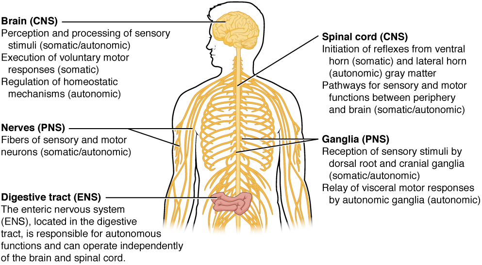
There is another division of the nervous system that describes functional responses. The enteric nervous system (ENS) is responsible for controlling the smooth muscle and glandular tissue in your digestive system. It is a large part of the PNS, and is not dependent on the CNS. It is sometimes valid, however, to consider the enteric system to be a part of the autonomic system because the neural structures that make up the enteric system are a component of the autonomic output that regulates digestion. There are some differences between the two, but for our purposes here there will be a good bit of overlap. See Figure 8.13 for examples of where these divisions of the nervous system can be found.
Functions of the Cerebral Cortex
The cerebrum is the seat of many of the higher mental functions, such as memory and learning, language, and conscious perception, which are the subjects of subtests of the mental status exam. The cerebral cortex is the thin layer of gray matter on the outside of the cerebrum. It is approximately a millimeter thick in most regions and highly folded to fit within the limited space of the cranial vault. These higher functions are distributed across various regions of the cortex, and specific locations can be said to be responsible for particular functions. There is a limited set of regions, for example, that are involved in language function, and they can be subdivided on the basis of the particular part of language function that each governs.
Cognitive Abilities
Assessment of cerebral functions is directed at cognitive abilities. The abilities assessed through the mental status exam can be separated into four groups: orientation and memory, language and speech, sensorium, and judgment and abstract reasoning.
Orientation and Memory
Orientation is the patient’s awareness of his or her immediate circumstances. It is awareness of time, not in terms of the clock but of the date and what is occurring around the patient. It is awareness of place, such that a patient should know where he or she is and why. It is also awareness of who the patient is—recognizing personal identity and being able to relate that to the examiner. The initial tests of orientation are based on the questions, “Do you know what the date is?” or “Do you know where you are?” or “What is your name?” Further understanding of a patient’s awareness of orientation can come from questions that address remote memory, such as “Who is the President of the United States?”, or asking what happened on a specific date.
Memory is largely a function of the temporal lobe, along with structures beneath the cerebral cortex such as the hippocampus and the amygdala. The storage of memory requires these structures of the medial temporal lobe. A famous case of a man who had both medial temporal lobes removed to treat intractable epilepsy provided insight into the relationship between the structures of the brain and the function of memory.
The prefrontal cortex can also be tested for the ability to organize information. In one subtest of the mental status exam called set generation, the patient is asked to generate a list of words that all start with the same letter, but not to include proper nouns or names. The expectation is that a person can generate such a list of at least 10 words within 1 minute. Many people can likely do this much more quickly, but the standard separates the accepted normal from those with compromised prefrontal cortices.
Language and Speech
Language is, arguably, a very human aspect of neurological function. There are certainly strides being made in understanding communication in other species, but much of what makes the human experience seemingly unique is its basis in language. Any understanding of our species is necessarily reflective, as suggested by the question “What am I?” And the fundamental answer to this question is suggested by the famous quote by René Descartes, “Cogito Ergo Sum” (translated from Latin as “I think, therefore I am”). Formulating an understanding of yourself is largely describing who you are to yourself. It is a confusing topic to delve into, but language is certainly at the core of what it means to be self-aware.
The neurological exam has two specific subtests that address language. One measures the ability of the patient to understand language by asking them to follow a set of instructions to perform an action, such as “touch your right finger to your left elbow and then to your right knee.” Another subtest assesses the fluency and coherency of language by having the patient generate descriptions of objects or scenes depicted in drawings, and by reciting sentences or explaining a written passage.
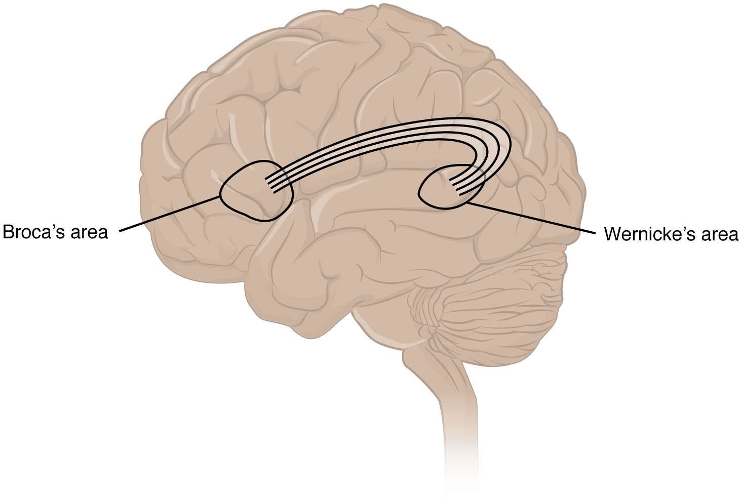
An important example of multimodal integrative areas is associated with language function (see Figure 8.14). Adjacent to the auditory association cortex, at the end of the lateral sulcus just anterior to the visual cortex, is Wernicke’s area. In the lateral aspect of the frontal lobe, just anterior to the region of the motor cortex associated with the head and neck is Broca’s area. Both regions were originally described on the basis of losses of speech and language, which is called aphasia. The aphasia associated with Broca’s area is known as expressive aphasia, which means that speech production is compromised. This type of aphasia is often described as non-fluency because the ability to say some words leads to broken or halting speech. Grammar can also appear to be lost. The aphasia associated with Wernicke’s area is known as receptive aphasia, which is not a loss of speech production but a loss of understanding of content. Patients, after recovering from acute forms of this aphasia, report not being able to understand what is said to them or what they are saying themselves, but they often cannot keep from talking.
The two regions are connected by white matter tracts that run between the posterior temporal lobe and the lateral aspect of the frontal lobe. Conduction aphasia associated with damage to this connection refers to the problem of connecting the understanding of language to the production of speech. This is a very rare condition but is likely to present as an inability to faithfully repeat spoken language.
Sensorium
Those parts of the brain involved in the reception and interpretation of sensory stimuli are referred to collectively as the sensorium. The cerebral cortex has several regions that are necessary for sensory perception. Several of the subtests can reveal activity associated with these sensory modalities, such as being able to hear a question or see a picture. Two subtests assess specific functions of these cortical areas.
The first is praxis, a practical exercise in which the patient performs a task completely on the basis of verbal description without any demonstration from the examiner. The second subtest for sensory perception is gnosis, which involves two tasks. The first task, known as stereognosis, involves the naming of objects strictly on the basis of the somatosensory information that comes from manipulating them. The patient keeps their eyes closed and is given a common object, such as a coin, that they have to identify. The patient should be able to indicate the particular type of coin, such as a dime versus a penny, or a nickel versus a quarter, on the basis of the sensory cues involved. For example, the size, thickness, or weight of the coin may be an indication, or to differentiate the pairs of coins suggested here, the smooth or corrugated edge of the coin will correspond to the particular denomination. The second task, graphesthesia, is to recognize numbers or letters written on the palm with a dull pointer, such as a pen cap.
Judgment and Abstract Reasoning
The prefrontal cortex is responsible for the functions responsible for planning and making decisions. In the mental status exam, the subtest that assesses judgment and reasoning is directed at three aspects of frontal lobe function. First, the examiner asks questions about problem-solving, such as “If you see a house on fire, what would you do?” The patient is also asked to interpret common proverbs, such as “Don’t look a gift horse in the mouth.” Additionally, pairs of words are compared for similarities, such as apple and orange, or lamp and cabinet.
Everyday Connections
Left Brain, Right Brain
Popular media often refer to right-brained and left-brained people, as if the brain were two independent halves that work differently for different people. This is a popular misinterpretation of an important neurological phenomenon. As an extreme measure to deal with a debilitating condition, the corpus callosum may be sectioned to overcome intractable epilepsy. When the connections between the two cerebral hemispheres are cut, interesting effects can be observed.
The reason for this is that the language functions of the cerebral cortex are localized to the left hemisphere in 95% of the population. Additionally, the left hemisphere is connected to the right side of the body through the corticospinal tract and the ascending tracts of the spinal cord. Motor commands from the precentral gyrus control the opposite side of the body, whereas sensory information processed by the postcentral gyrus is received from the opposite side of the body. For a verbal command to initiate movement of the right arm and hand, the left side of the brain needs to be connected by the corpus callosum. Language is processed in the left side of the brain and directly influences the left brain and right arm motor functions but is sent to influence the right brain and left arm motor functions through the corpus callosum. Likewise, the left-handed sensory perception of what is in the left pocket travels across the corpus callosum from the right brain, so no verbal report on those contents would be possible if the hand happened to be in the pocket.
People who have had their corpus callosum cut can perform two independent tasks at the same time because the lines of communication between the right and left sides of their brains have been removed. Whereas a person with an intact corpus callosum cannot overcome the dominance of one hemisphere over the other, this patient can. If the left cerebral hemisphere is dominant in the majority of people, why would right-handedness be most common?
Common Abbreviations for the Nervous System
Disease and Disorders
Neurodegenerative Diseases – Alzheimer’s disease, Parkinson’s disease, Amyotrophic Lateral Sclerosis (ALS), Multiple sclerosis (MS)
A class of disorders that affect the nervous system are the neurodegenerative diseases: Alzheimer’s disease, Parkinson’s disease, Huntington’s disease, amyotrophic lateral sclerosis (ALS), Creutzfeldt–Jakob disease, multiple sclerosis (MS), and other disorders that are the result of nervous tissue degeneration. In diseases like Alzheimer’s, Parkinson’s, or ALS, neurons die; in diseases like MS, myelin is affected. Some of these disorders affect motor function, and others present with dementia. Some are the result of genetics, such as Huntington’s disease, or the result of autoimmunity, such as MS; others are not entirely understood, such as Alzheimer’s and Parkinson’s diseases.
Several diseases can result from the demyelination of axons. The causes of these diseases are not the same; some have genetic causes, some are caused by pathogens, and others are the result of autoimmune disorders. Though the causes are varied, the results are largely similar. The myelin insulation of axons is compromised, making electrical signaling slower.
Multiple sclerosis (MS) is one such disease. It is an example of an autoimmune disease. The antibodies produced by lymphocytes (a type of white blood cell) mark myelin as something that should not be in the body. This causes inflammation and the destruction of the myelin in the central nervous system. As the insulation around the axons is destroyed by the disease, scarring becomes obvious.
Guillain-Barre (pronounced gee-YAN bah-RAY) syndrome is an example of a demyelinating disease of the peripheral nervous system. It is also the result of an autoimmune reaction, but the inflammation is in peripheral nerves. Sensory symptoms or motor deficits are common, and autonomic failures can lead to changes in the heart rhythm or a drop in blood pressure, especially when standing, which causes dizziness.
Other Nerve Disorders
Infection, trauma, and congenital disorders can all lead to significant signs, as identified through the neurological exam. It is important to differentiate between an acute event, such as stroke, and a chronic or global condition such as blunt force trauma. Responses seen in the neurological exam can help. A loss of language function observed in all its aspects is more likely a global event as opposed to a discrete loss of one function, such as not being able to say certain types of words. A concern, however, is that a specific function—such as controlling the muscles of speech—may mask other language functions. The various subtests within the mental status exam can address these finer points and help clarify the underlying cause of the neurological loss.
Stroke
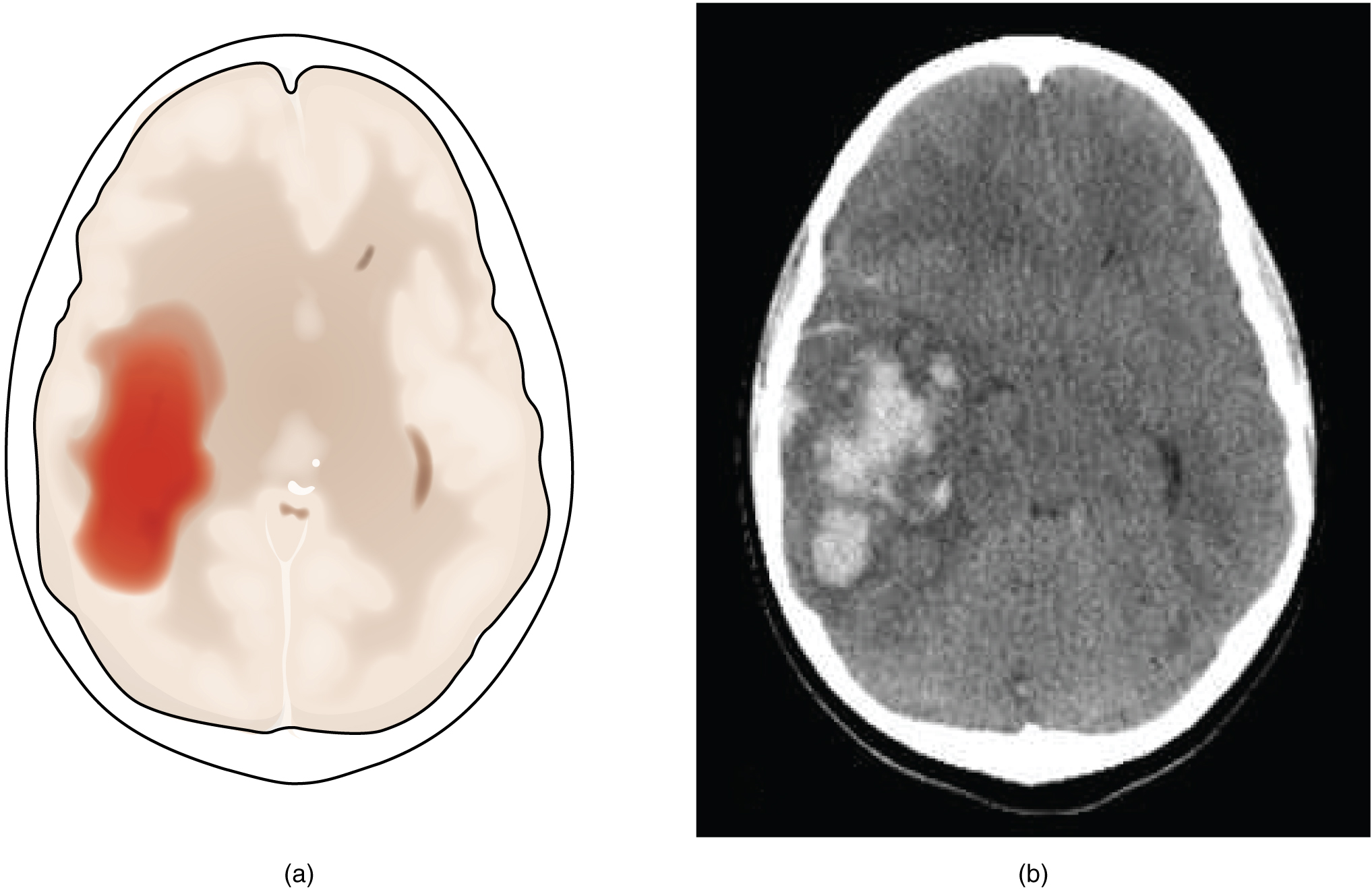
Damage to the nervous system can be limited to individual structures or can be distributed across broad areas of the brain and spinal cord. Localized, limited injury to the nervous system is most often the result of circulatory problems. The loss of blood flow to part of the brain is known as a stroke, or a cerebrovascular accident (CVA). There are two main types of stroke, depending on how the blood supply is compromised: ischemic and hemorrhagic. An ischemic stroke is the loss of blood flow to an area because vessels are blocked or narrowed. This is often caused by an embolus, which may be a blood clot or fat deposit. Ischemia may also be the result of thickening of the blood vessel wall, or a drop in blood volume in the brain known as hypovolemia. A hemorrhagic stroke is bleeding into the brain because of a damaged blood vessel. Accumulated blood fills a region of the cranial vault and presses against the tissue in the brain (see Figure 8.15).
Cerebral Palsy
Cerebral palsy (CP) is caused by an interruption to the normal development of a person’s brain, leading to weakness with muscles. Depending on the area of the brain that is affected, signs and symptoms will vary in the type and severity between individuals. Balance and coordination are often challenging due to the inability to control muscles (Centers for Disease Control and Prevention, n.d.-a). To learn more about cerebral palsy, please visit the Centers for Disease Control and Prevention.
Traumatic Brain Injury (TBI)
According to the Centers for Disease Control and Prevention, about 166 people in the United States died each day from a traumatic brain injury in 2019. Brain injuries range from mild to severe and include concussions. TBI can be caused by falls, automobile accidents, assaults, and firearm-related suicide (Centers for Disease Control and Prevention, n.d.-b). To learn more about TBI, please visit the Centers for Disease Control and Prevention.
Practice Medical Terms in Context
Medical Specialties
Neurologist
Neurologists are medical doctors who complete specialized training in the prevention, diagnosis, and treatment of disorders and conditions related to the brain and nervous system (Bureau of Labor Statistics, 2021). For more details visit the American Academy of Neurology.
Procedures Related to the Nervous System
Lumbar Puncture
Electromyography (EMG)
Electromyography (EMG) is a procedure that assesses the electrical signals muscles send while at rest and when they are used. During the test, a needle electrode is placed into the muscle, and a machine records the muscle activity. EMG can be used to diagnose myasthenia gravis, muscular dystrophy, and other conditions affecting the muscles (MedlinePlus, 2021a). To learn more, please visit the Medline Plus web page on electromyography.
Electroencephalogram (EEG)
With electrodes applied to your scalp, an electroencephalogram (EEG) measures electrical activity in the brain. It’s used to help diagnose conditions of the brain, including seizures, altered mental status, and hemorrhage. (Rayi & Murr, 2021). For more information, please visit the Mayo Clinic’s web page on electroencephalograms.
Practice Terms Related to the Nervous System
Nervous System Vocabulary
- Afferent nerves
Nerves that carry sensory signals (nerve impulses) toward the central nervous from the periphery.
Aphasia
Loss of language function.
Arachnoid mater
Middle layer of the meninges named for the spider-web–like trabeculae that extend between it and the pia mater.
Astrocyte
Glial cell type of the central nervous system that provides support for neurons and maintains the blood-brain barrier.
Autonomic nervous system (ANS)
Functional division of the nervous system that is responsible for homeostatic reflexes that coordinate control of cardiac and smooth muscle, as well as glandular tissue.
Axon
Single process of the neuron that carries an electrical signal (action potential) away from the cell body toward a target cell.
Axon hillock
Tapering of the neuron cell body that gives rise to the axon.
Axon segment
Single stretch of the axon insulated by myelin and bounded by nodes of Ranvier at either end (except for the first, which is after the initial segment, and the last, which is followed by the axon terminal).
Axon terminal
End of the axon, where there are usually several branches extending toward the target cell.
Axoplasm
Cytoplasm of an axon, which is different in composition than the cytoplasm of the neuronal cell body.
Babinski sign
Dorsiflexion of the foot with extension and splaying of the toes in response to the plantar reflex, normally suppressed by corticospinal input.
Bipolar
Shape of a neuron with two processes extending from the neuron cell body—the axon and one dendrite.
Blood-brain barrier (BBB)
Physiological barrier between the circulatory system and the central nervous system that establishes a privileged blood supply, restricting the flow of substances into the central nervous system.
Brain
The large organ of the central nervous system composed of white and gray matter, contained within the cranium and continuous with the spinal cord.
Brain stem
Region of the adult brain that includes the midbrain, pons, and medulla oblongata and develops from the mesencephalon, metencephalon, and myelencephalon of the embryonic brain.
Broca’s area
Region of the frontal lobe associated with the motor commands necessary for speech production.
Brodmann’s areas
Mapping of regions of the cerebral cortex based on microscopic anatomy that relates specific areas to functional differences, as described by Brodmann in the early 1900s.
Cauda equina
Bundle of spinal nerve roots that descend from the lower spinal cord below the first lumbar vertebra and lie within the vertebral cavity; has the appearance of a horse’s tail.
Caudate
Nucleus deep in the cerebrum that is part of the basal nuclei; along with the putamen, it is part of the striatum.
Central nervous system (CNS)
Anatomical division of the nervous system located within the cranial and vertebral cavities, namely the brain and spinal cord.
Central sulcus
Surface landmark of the cerebral cortex that marks the boundary between the frontal and parietal lobes.
Cephalgia
Pain in the head.
Cerebellum
Region of the adult brain connected primarily to the pons that developed from the metencephalon (along with the pons) and is largely responsible for comparing information from the cerebrum with sensory feedback from the periphery through the spinal cord.
Cerebral angiography
Process of recording the blood vessels of the cerebrum.
Cerebral cortex
Outer gray matter covering the forebrain, marked by wrinkles and folds known as gyri and sulci.
Cerebrum
Region of the adult brain that develops from the telencephalon and is responsible for higher neurological functions such as memory, emotion, and consciousness.
Cerebral hemisphere
One half of the bilaterally symmetrical cerebrum.
Cerebrospinal fluid (CSF)
A colorless fluid produced by the brain that cushions the brain and spinal cord within the posterior (dorsal) cavity.
Cerebral thrombosis
Formation of a blood clot in a blood vessel within the skull.
Choroid plexus
Specialized structure containing ependymal cells that line blood capillaries and filter blood to produce cerebrospinal fluid in the four ventricles of the brain.
Corpus callosum
Large white matter structure that connects the right and left cerebral hemispheres.
Dendrite
One of many branchlike processes that extends from the neuron cell body and functions as a contact for incoming signals (synapses) from other neurons or sensory cells.
Descending tract
Central nervous system fibers carrying motor commands from the brain to the spinal cord or periphery.
Diencephalon
Region of the adult brain that retains its name from embryonic development and includes the thalamus and hypothalamus.
Direct pathway
Connections within the basal nuclei from the striatum to the globus pallidus internal segment and substantia nigra pars reticulata that disinhibit the thalamus to increase cortical control of movement.
Dorsal (posterior) nerve root
Axons entering the posterior horn of the spinal cord.
Dura mater
Tough, fibrous, outer layer of the meninges that is attached to the inner surface of the cranium and vertebral column and surrounds the entire central nervous system.
Efferent nerves
Nerve tissue that carries impulses away from the CNS towards the peripheral that result in motor response (movement).
Electroencephalogram
The record of electrical activity of the brain.
Electroencephalography
Process of recording the electrical activity of the brain.
Embolus
An obstruction such as a blood clot or plaque that blocks the flow of blood in an artery or vein.
Encephalitis
Inflammation of the tissues of the brain.
Encephalomalacia
Softening of the tissues of the brain.
Enteric nervous system (ENS)
Neural tissue associated with the digestive system that is responsible for nervous control through autonomic connections.
Ependymal cell
Glial cell type in the central nervous system responsible for producing cerebrospinal fluid.
Epithalamus
Region of the diencephalon containing the pineal gland.
Foramen magnum
Large opening in the occipital bone of the skull through which the spinal cord emerges and the vertebral arteries enter the cranium.
Frontal lobe
Region of the cerebral cortex directly beneath the frontal bone of the cranium.
Ganglion
Localized collection of neuron cell bodies in the peripheral nervous system.
Ganglionectomy
Excision of a ganglion.
Glial cell
One of the various types of neural tissue cells responsible for maintenance of the tissue, and largely responsible for supporting neurons.
Glioblastoma
A central nervous system tumor composed of developing glial tissue.
Glioma
A tumor that begins in the glial tissue.
Gray matter
Regions of the nervous system containing cell bodies of neurons with few or no myelinated axons; actually may be more pink or tan in color, but called gray in contrast to white matter.
Gyrus
Ridge formed by convolutions on the surface of the cerebrum or cerebellum.
Hemiplegia
Paralysis on one side of the body.
Hemorrhagic stroke
Disruption of blood flow to the brain caused by bleeding within the cranial vault.
Hydrocephalus
The abnormal buildup of cerebrospinal fluid in the ventricles of the brain.
Hyperesthesia
Increased sensitivity to stimuli.
Hypothalamus
A region of the forebrain below the thalamus; has function in both the autonomic and endocrine systems and regulates homeostasis.
Ischemic stroke
Disruption of blood flow to the brain because blood cannot flow through blood vessels as a result of a blockage or narrowing of the vessel.
Integration
Nervous system function that combines sensory perceptions and higher cognitive functions (memories, learning, emotion, etc.) to produce a response.
Initial segment
First part of the axon as it emerges from the axon hillock, where the electrical signals known as action potentials are generated.
Longitudinal fissure
A large separation along the midline between the two cerebral hemispheres.
Lumbar puncture
Procedure used to withdraw cerebrospinal fluid from the lower lumbar region of the vertebral column.
Medulla oblongata
A part of the brain stem responsible for control of heart rate and breathing.
Meninges
The membranes that surround the central nervous system.
Meningioma
A tumor of the meninges.
Meningitis
Inflammation of the meninges, the tough membranes that surround the central nervous system.
Meningocele
Protrusion of the meninges.
Meningomyelocele
Protrusion of the meninges and spinal cord.
Microglia
Smaller than most of the other glial cells; they ingest and digest cells or pathogens that cause disease.
Midbrain
A portion of the brainstem, positioned above the pons, also called mesencephalon, that assists in motor reflexes associated with visual, auditory, and somatosensory stimuli.
Mononeuropathy
Disease affecting a single peripheral nerve.
Motor nerves
Peripheral, efferent, myelinated nerve tissue that stimulates muscle contraction.
Multipolar
Shape of a neuron that has multiple processes—the axon and two or more dendrites.
Myelin sheath
Lipid-rich layer of insulation that surrounds an axon, formed by oligodendrocytes in the central nervous system and Schwann cells in the peripheral nervous system; facilitates the transmission of electrical signals.
Nerves
Bundle of fibers that receives and sends messages between the body and the brain.
Neuralgia
Pain of the peripheral or cranial nerves.
Neuritis
Inflammation of a peripheral or cranial nerve.
Neuroglia
- Supportive tissue of the nervous system, including the network of branched cells in the central nervous system (astrocytes, microglia, and oligodendrocytes) and the supporting cells of the peripheral nervous system (Schwann cells and satellite cells), also called glia.
Neurologist
A doctor who has special training in diagnosing and treating disorders of the nervous system.
Neurology
A medical specialty concerned with the study of the structures, functions, and diseases of the nervous system.
Neuroma
Tumor made up of nerve cells.
Neuron
Cells that propagate information via electrochemical impulses.
Neuropathy
A nerve problem that causes pain, numbness, tingling, swelling, or muscle weakness in different parts of the body.
Neurotransmitters
Chemicals that are made by nerve cells and used to communicate with other cells, including other nerve cells and muscle cells.
Node of Ranvier
Gap between two myelinated regions of an axon, allowing for strengthening of the electrical signal as it propagates down the axon.
Nucleus
The cell’s central organelle, which contains the cell’s DNA.
Occipital lobe
Region of the cerebral cortex directly beneath the occipital bone of the cranium.
Olfaction
The sense of smell.
Oligodendrocyte
Glial cell type in the central nervous system that provides the myelin insulation for axons in tracts.
Paresis
Partial paralysis wherein there is still some control of the muscles.
Paresthesia
Abnormal sensation in the extremities.
Parietal lobe
Region of the cerebral cortex directly beneath the parietal bone of the cranium.
Peripheral nervous system (PNS)
All nervous tissue that is outside of the brain and spinal cord.
Pia mater
Thin, innermost membrane of the meninges that directly covers the surface of the central nervous system.
Poliomyelitis
Acute infection by the poliovirus, especially of the motor neurons in the spinal cord and brainstem.
Polyneuritis
Inflammation of several peripheral nerves at the same time.
Polyneuropathy
Disease of multiple peripheral nerves at the same time.
Pons
The main connection between the cerebellum and the brain stem. It is responsible for regulating several crucial functions, including the cardiovascular and respiratory systems.
Process
In cells, an extension of a cell body; in the case of neurons, this includes the axon and dendrites.
Psychiatrist
A medical doctor who specializes in neuroscience and diagnoses and treats mental disorders.
Psychiatry
The medical science that deals with the origin, diagnosis, prevention, and treatment of mental disorders.
Psychologist
A specialist who can talk with patients and their families about emotional and personal matters.
Psychology
The study of how the mind works and how thoughts and feelings affect behavior.
Psychosis
A severe mental disorder in which a person loses the ability to recognize reality or relate to others.
Quadriplegia
Paralysis of all four limbs.
Radiculopathy
Disease of the nerve roots.
Response
Nervous system function that causes a target tissue (muscle or gland) to produce an event as a consequence to stimuli.
Rhizotomy
Incision into a nerve root.
Satellite cell
Glial cell type in the peripheral nervous system that provides support for neurons in the ganglia.
Schwann cell
Glial cell type in the peripheral nervous system that provides the myelin insulation for axons in nerves.
Sensation
Nervous system function that receives information from the environment and translates it into the electrical signals of nervous tissue.
Soma
In neurons, that portion of the cell that contains the nucleus; the cell body, as opposed to the cell processes (axons and dendrites).
Somatic nervous system (SNS)
Functional division of the nervous system that is concerned with conscious perception, voluntary movement, and skeletal muscle reflexes.
Spinal cord
Organ of the central nervous system found within the vertebral cavity and connected with the periphery through spinal nerves; mediates reflex behaviors.
Stimulus
An event in the external or internal environment that registers as activity in a sensory neuron.
Stroke
Loss of neurological function caused by an interruption of blood flow to a region of the central nervous system, also called cerebrovascular accident (CVA).
Subarachnoid space
Space between the arachnoid mater and pia mater that contains CSF and the fibrous connections of the arachnoid trabeculae.
Subdural hematoma
Accumulation of blood in the subdural space.
Sulcus
Groove formed by convolutions in the surface of the cerebral cortex.
Synapse
Narrow junction across which a chemical signal passes from neuron to the next, initiating a new electrical signal in the target cell.
Synaptic end bulb
Swelling at the end of an axon where neurotransmitter molecules are released onto a target cell across a synapse.
Sympathetic nervous system (SNS)
The division of the nervous system involved in our fight-or-flight responses. It continuously monitors body temperature and initiates appropriate motor responses.
Temporal lobe
Region of the cerebral cortex directly beneath the temporal bone of the cranium.
Thalamus
Major region of the diencephalon that is responsible for relaying information between the cerebrum and the hindbrain, spinal cord, and periphery.
Tract
Bundle of axons in the central nervous system having the same function and point of origin.
Transient ischemic attack (TIA)
Temporary disruption of blood flow to the brain in which symptoms occur rapidly but last only a short time.
Unipolar
Shape of a neuron which has only one process that includes both the axon and dendrite.
Ventricle
Central cavity within the brain where cerebrospinal fluid is produced and circulates.
Wernicke’s area
Region at the posterior end of the lateral sulcus in which speech comprehension is localized.
White matter
Regions of the nervous system containing mostly myelinated axons, making the tissue appear white because of the high lipid content of myelin.
- Supportive tissue of the nervous system, including the network of branched cells in the central nervous system (astrocytes, microglia, and oligodendrocytes) and the supporting cells of the peripheral nervous system (Schwann cells and satellite cells), also called glia.
Test Yourself
References
Bureau of Labor Statistics. (2021). Physicians and surgeons. In Occupational outlook handbook. U.S. Department of Labor. https://www.bls.gov/ooh/healthcare/physicians-and-surgeons.htm
Centers for Disease Control and Prevention. (n.d.-a). What is cerebral palsy? https://www.cdc.gov/ncbddd/cp/facts.html
Centers for Disease Control and Prevention. (n.d.-b). Get the facts about TBI. https://www.cdc.gov/traumaticbraininjury/get_the_facts.html
ClinicalInfo. (n.d.). Spinal tap. National Institute of Health Office of AIDS Research, U.S. Department of Health and Human Services. https://clinicalinfo.hiv.gov/en/glossary/spinal-tap
CrashCourse. (2015, February 23). The nervous system, part 1: Crash course A&P #8 [Video]. YouTube. https://www.youtube.com/watch?v=qPix_X-9t7E
MedlinePlus. (2021a). Electromyography (EMG) and nerve conduction studies. U.S. Library of Medicine, U.S. Department of Health and Human Services. https://medlineplus.gov/lab-tests/electromyography-emg-and-nerve-conduction-studies
Rayi, A., & Murr, N. (2021). Electroencephalogram. In StatPearls [Internet]. https://www.ncbi.nlm.nih.gov/books/NBK563295/
Image Descriptions
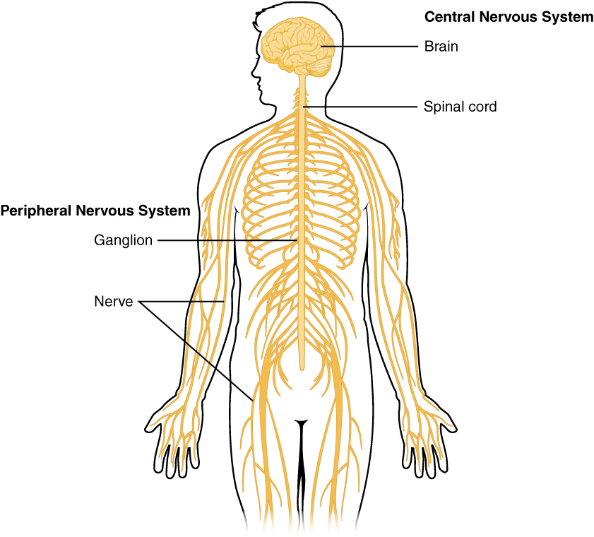
Figure 8.1 image description: This diagram shows a silhouette of a human highlighting the nervous system. The central nervous system is composed of the brain and spinal cord. The brain is a large mass of ridged and striated tissue within the head. The spinal cord extends down from the brain and travels through the torso, ending in the pelvis. Pairs of enlarged nervous tissue, labeled ganglia, flank the spinal cord as it travels through the rib area. The ganglia are part of the peripheral nervous system, along with the many thread-like nerves that radiate from the spinal cord and ganglia through the arms, abdomen, and legs. [Return to Figure 8.1].
Figure 8.2 image description: This photo shows an enlarged view of the dorsal side of a human brain. The right side of the occipital lobe has been shaved to reveal the white and gray matter beneath the surface blood vessels. The white matter branches through the shaved section like the limbs of a tree. The gray matter branches and curves on the outside of the white matter, creating a buffer between the outer edges of the occipital lobe and the internal white matter. [Return to Figure 8.2].
Figure 8.3 image description: This figure shows the lateral view on the left panel and the anterior view on the right panel of the brain. The major parts including the cerebrum are labeled. Lateral view labels (clockwise from top) read: cerebrum, cerebral cortex, corpus callosum (located on the interior of the brain). Anterior view labels indicate the right and left hemispheres and the longitudinal fissure between them. [Return to Figure 8.3].
Figure 8.4 image description: This figure shows the lateral view of the brain and the major lobes are labeled. From the front of the brain (left) labels read: frontal lobe, precentral gyrus, central sulcus, postcentral gyrus, parietal lobe, lateral sulcus, occipital lobe, temporal lobe. [Return to Figure 8.4].
Figure 8.5 image description: This figure shows the location of the thalamus, hypothalamus, and pituitary gland in the brain. Each part is labeled respectively. The thalamus is located in the midsection of the brain. The hypothalamus is located below the thalamus and the pituitary gland below that. [Return to Figure 8.5].
Figure 8.6 image description: This figure shows the location of the midbrain, pons, and the medulla in the brain that make up the brainstem. The midbrain is located at the top, the pons is located beneath that, and the medulla is the lowest most point of the brain stem. [Return to Figure 8.6].
Figure 8.7 image description: This figure shows the location of the cerebellum in the brain which is located on the posterior surface of the brain stem. Labels read (top, left): pons, inferior olive, (top, right) cerebellum, deep cerebellar white matter (arbor vitae). In the top panel, a lateral view labels the location of the cerebellum and the deep cerebellar white matter. In the bottom panel, a photograph of a brain, with the cerebellum in pink is shown. [Return to Figure 8.7].
Figure 8.8 image description: This illustration shows the anatomy of a neuron. The neuron has a very irregular cell body (soma) containing a purple nucleus. There are six projections protruding from the top, bottom, and left sides of the cell body. Each of the projections branches many times, forming small, tree-shaped structures protruding from the cell body. The right side of the cell body tapers into a long cord called the axon. The axon is insulated by segments of myelin sheath, which resemble a semitransparent toilet paper roll wound around the axon. The myelin sheath is not continuous but is separated into equally spaced segments. The bare axon segments between the sheath segments are called nodes of Ranvier. An oligodendrocyte is reaching its two arm-like projections onto two myelin sheath segments. The axon branches many times at its end, where it connects to the dendrites of another neuron. Each connection between an axon branch and a dendrite is called a synapse. The cell membrane completely surrounds the cell body, dendrites, and axon. The axon of another nerve is seen in the upper left of the diagram connecting with the dendrites of the central neuron. [Return to Figure 8.8].
Figure 8.9 image description: Three illustrations show some of the possible shapes that neurons can take. In the unipolar neuron, the dendrite enters from the left and merges with the axon into a common pathway, which is connected to the cell body. The axon leaves the cell body through the common pathway, the branches off to the right, in the opposite direction as the dendrite. Therefore, this neuron is T-shaped. In the bipolar neuron, the dendrite enters into the left side of the cell body while the axon emerges from the opposite (right) side. In a multipolar neuron, multiple dendrites enter the cell body. The only part of the cell body that does not have dendrites is the part that elongates into the axon. [Return to Figure 8.9].
Figure 8.10 image description: This diagram contains three black and white drawings of more specialized nerve cells. Part A shows a pyramidal cell of the cerebral cortex, which has two, long, nerve tracts attached to the top and bottom of the cell body. However, the cell body also has many short dendrites projecting out a short distance from the cell body. Part B shows a Purkinje cell of the cerebellar cortex. This cell has a single, long, nerve tract entering the bottom of the cell body. Two large nerve tracts leave the top of the cell body but immediately branch many times to form a large web of nerve fibers. Therefore, the Purkinje cell somewhat resembles a shrub or coral in shape. Part C shows the olfactory cells in the olfactory epithelium and olfactory bulbs. It contains several cell groups linked together. At the bottom, there is a row of olfactory epithelial cells that are tightly packed, side-by-side, somewhat resembling the slats on a fence. There are six neurons embedded in this epithelium. Each neuron connects to the epithelium through branching nerve fibers projecting from the bottom of their cell bodies. A single nerve fiber projects from the top of each neuron and synapses with nerve fibers from the neurons above. These upper neurons are cross-shaped, with one nerve fiber projecting from the bottom, top, right and left sides. The upper cells synapse with the epithelial nerve cells using the nerve tract projecting from the bottom of their cell body. The nerve tract projecting from the top continues the pathway, making a ninety-degree turn to the right and continuing to the right border of the image. [Return to Figure 8.10].
Figure 8.11 image description: This diagram shows several types of nervous system cells associated with two multipolar neurons. Astrocytes are star shaped-cells with many dendrite-like projections but no axon. They are connected with the multipolar neurons and other cells in the diagram through their dendrite-like projections. Ependymal cells have a teardrop-shaped cell body and a long tail that branches several times before connecting with astrocytes and the multipolar neuron. Microglial cells are small cells with rectangular bodies and many dendrite-like projections stemming from their shorter sides. The projections are so extensive that they give the microglial cell a fuzzy appearance. The oligodendrocytes have circular cell bodies with four dendrite-like projections. Each projection is connected to a segment of myelin sheath on the axons of the multipolar neurons. The oligodendrocytes are the same color as the myelin sheath segment and are adding layers to the sheath using their projections. [Return to Figure 8.11].
Figure 8.12 image description: This diagram shows a collection of PNS glial cells. The largest cell is a unipolar peripheral ganglionic neuron which has a common nerve tract projecting from the bottom of its cell body. The common nerve tract then splits into the axon, going off to the left, and the dendrite, going off to the right. The cell body of the neuron is covered with several satellite cells that are irregular, flattened, and take on the appearance of fried eggs. Schwann cells wrap around each myelin sheath segment on the axon, with their nucleus creating a small bump on each segment. [Return to Figure 8.12].
Figure 8.13 image description: A silhouette of a human with only the brain, spinal cord, PNS ganglia, nerves, and a section of the digestive tract visible. The brain, which is part of the CNS, is the area of perception and processing of sensory stimuli (somatic/autonomic), the execution of voluntary motor responses (somatic), and the regulation of homeostatic mechanisms (autonomic). The spinal cord, which is part of the CNS, is the area where reflexes are initiated. The gray matter of the ventral horn initiates somatic reflexes while the gray matter of the lateral horn initiates autonomic reflexes. The spinal cord is also the somatic and autonomic pathway for sensory and motor functions between the PNS and the brain. The nerves, which are part of the PNS, are the fibers of sensory and motor neurons, which can be either somatic or autonomic. The ganglia, which are part of the PNS, are the areas for the reception of somatic and autonomic sensory stimuli. These are received by the dorsal root ganglia and cranial ganglia. The autonomic ganglia are also the relay for visceral motor responses. The digestive tract is part of the enteric nervous system, the ENS, which is located in the digestive tract and is responsible for the autonomous function. The ENS can operate independently of the brain and spinal cord. [Return to Figure 8.13].
Figure 8.14 image description: An illustration of the brain with Broca’s area and Wernicke’s area identified. Broca’s area is located in the lateral aspect of the frontal lobe. Wernicke’s area is found at the end of the lateral sulcus just anterior to the visual cortex. The two are connected by white matter tracts between the posterior temporal love and lateral aspect of the frontal lobe. Both areas are associated with the loss of speech and language. Expressive aphasia is associated with Broca’s area. Receptive aphasia is associated with Wernicke’s area. [Return to Figure 8.14].
Figure 8.15 image description: The left panel of this figure shows an image of the brain with a region in red. This red region indicates a hemorrhage associated with a stroke. The right panel shows a hemorrhage as it might appear on a CT scan. [Return to Figure 8.15].
An instrument that is used to look at cells and other small objects that cannot be seen with the eye alone (National Cancer Institute, n.d.)
The study of electrical properties of cells and tissues (National Library of Medicine, 2021)
Anatomical division of the nervous system located within the cranial and vertebral cavities, namely the brain and spinal cord (Betts et al., 2013)
All nervous tissue that is outside of the brain and spinal cord (Betts et al., 2013)
Region of the adult brain that develops from the telencephalon and is responsible for higher neurological functions such as memory, emotion, and consciousness (Betts et al., 2013)
A region of the forebrain below the thalamus; has function in both the autonomic and endocrine systems and regulates homeostasis (Betts et al., 2013)
