
7 Muscular System
Learning Objectives
- Examine the anatomy of the muscular system
- Determine the main functions of the muscular system
- Differentiate the medical terms of the muscular system and common abbreviations
- Discover common diseases, disorders, and procedures related to the muscular system
- Recognize the medical specialties associated with the muscular system
Muscular System Word Parts
Click on prefixes, combining forms, and suffixes to reveal a list of word parts to memorize for the Muscular System.
Introduction to the Muscular System
When most people think of muscles, they think of the muscles that are visible just under the skin, particularly of the limbs. These are skeletal muscles, so-named because most of them move the skeleton, but there are two additional types of muscles: the smooth muscle and the cardiac muscle. The body has over 600 muscles which contribute significantly to the body’s weight.
Watch this video:
Media 7.1 Muscles, Part 2 – Organismal Level: Crash Course A&P #22 [Online video]. Copyright 2015 by CrashCourse.
Medical Terms Related to the Muscular System
Anatomy (Structures) of the Muscular System
Muscle is one of the four primary tissue types of the body, and it is made up of specialized cells called fibers. The body contains three types of muscle tissue: skeletal muscle, cardiac muscle, and smooth muscle (see Figure 7.1). All three muscle tissues have some properties in common; they all exhibit a quality called excitability as their plasma membranes can change their electrical states (from polarized to depolarized) and send an electrical wave called an action potential along the entire length of the membrane. Fascia is fibrous connective tissue that encloses muscles.
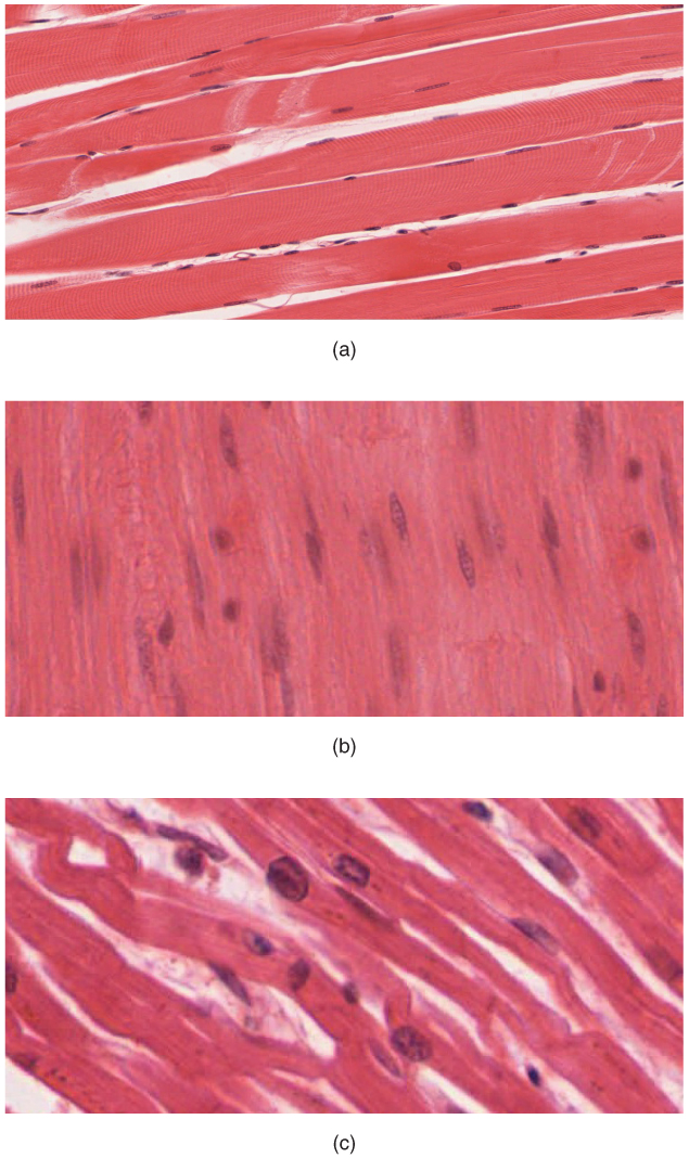
Three Types of Muscle Tissues
- Skeletal – closely associated with the skeletal system. Also known as striated muscles, they are responsible for voluntary muscle movement, such as swallowing, et cetera.
- Smooth – mainly associated with the walls of the internal organs. Also known as visceral muscles, they are responsible for involuntary muscle movement, such as breathing, et cetera.
- Cardiac – heart muscle or myocardium. Its appearance is similar to a skeletal muscle and is responsible for the pumping of blood. It gives the heart beat.
Skeletal Muscle
Skeletal muscles act not only to produce movement but also to stop movement, such as resisting gravity to maintain posture. Small, constant adjustments of the skeletal muscles are needed to hold a body upright or balanced in any position. Muscles also prevent excess movement of the bones and joints, maintaining skeletal stability and preventing skeletal structure damage or deformation.
Skeletal muscles are located throughout the body at the openings of internal tracts to control the movement of various substances. These muscles allow functions, such as swallowing, urination, and defecation, to be under voluntary control. Skeletal muscles also protect internal organs (particularly abdominal and pelvic organs) by acting as an external barrier or shield to external trauma and by supporting the weight of the organs.
Skeletal muscles contribute to the maintenance of homeostasis in the body by generating heat. This heat is very noticeable during exercise, when sustained muscle movement causes body temperature to rise, and in cases of extreme cold when shivering produces random skeletal muscle contractions to generate heat.
Did you know?
The gluteus maximus is the largest muscle and the heart is the hardest working muscle.
Smooth Muscle
Smooth muscle, so named because the cells do not have striations, is present in the walls of hollow organs like the urinary bladder, uterus, stomach, intestines, and in the walls of passageways, such as the arteries and veins of the circulatory system, and the tracts of the respiratory, urinary, and reproductive systems. Smooth muscle is also present in the eyes, where it functions to change the size of the iris and alter the shape of the lens; and in the skin where it causes hair to stand erect in response to cold temperature or fear.
Cardiac Muscle
Cardiac muscle tissue is only found in the heart. Highly coordinated contractions of cardiac muscle pump blood into the vessels of the circulatory system. Similar to skeletal muscle, cardiac muscle is striated and organized into sarcomeres, possessing the same banding organization as skeletal muscle (see Figure 7.1). Cardiac muscle fibers cells also are extensively branched and are connected to one another at their ends by intercalated discs. An intercalated disc allows the cardiac muscle cells to contract in a wave-like pattern so that the heart can work as a pump.
Concept Check
- Compare and contrast the three types of muscle tissues.
- Where in the body do you find each of the muscle types?
Physiology (Function) of the Muscular System
The main function of the muscular system is to assist with movement. Muscles work as antagonistic pairs. As one muscle contracts, the other muscle relaxes. This contraction pulls on the bones and assists with movement. Contraction is the shortening of the muscle fibers while relaxation lengthens the fibers. This sequence of relaxation and contraction is influenced by the nervous system.
Muscles also work to keep the posture of the body. This is done through muscle contraction where the trunk is kept straight either when sitting or standing.
Did you know?
If all the muscles in the jaw worked together, it could close the teeth with a force as great as 200 pounds on the molars (Science Reference Section, 2019).
Naming of Muscles
There are many nomenclatures for naming muscles. Some of these include:
- divisions – biceps, triceps, quadriceps
- size – maximus (largest), minimus (smallest)
- shape – deltoid (triangular), trapezius (trapezoid)
- action – flexor (to flex), adductor (towards midline of the body)
| EXAMPLE | WORD | LATIN ROOT 1 | LATIN ROOT 2 | MEANING | TRANSLATION |
| abductor digiti minimi | abductor | ab = away from | duct = to move | a muscle that moves away from | A muscle that moves the little finger or toe away |
| digiti | digitus = digit | n/a | refers to a finger or toe | ||
| minimi | minimus = mini, tiny | n/a | little | ||
| adductor digiti minimi | adductor | ad = to, toward | duct = to move | a muscle that moves towards | A muscle that moves the little finger or toe toward |
| digiti | digitus = digit | n/a | refers to a finger or toe | ||
| minimi | minimus = mini, tiny | n/a | little |
Common Abbreviations for the Muscular System
Many terms and phrases related to the muscle system are abbreviated. Learn these common abbreviations by expanding the list below.
Diseases and Disorders of the Muscle System
Duchenne Muscular Dystrophy
Duchenne Muscular Dystrophy (DMD) is caused by the inability of the body to make dystrophin (a muscle protein). This causes the muscles to become weak as the person ages. This disease primarily affects boys. Signs and symptoms typically present before the age of six and may include a delay of motor milestones and progressive weakness in the lower extremities and pelvis. Since all muscles are affected, the person will eventually require a wheelchair and assistance with breathing (National Human Genome Research Institute, 2013). To learn more, please visit the Genetic and Rare Diseases Information Center’s web page on Duchenne Muscular Dystrophy.
Cerebral Palsy
Cerebral palsy (CP) is caused by an interruption to the normal development of a person’s brain leading to weakness with muscles. Depending on the area of the brain that is affected, signs and symptoms will vary in the type and severity between individuals. Balance and coordination are often challenging due to the inability to control muscles (Centers for Disease Control and Prevention, n.d.). To learn more, please visit the Centers for Disease Control and Prevention’s web page on cerebral palsy.
Carpal Tunnel Syndrome
Carpal tunnel syndrome may present with pain, numbness, or weakness to the hand(s) caused by pressure on the median nerve. Some causes for this pressure are repetitive movements, trauma or injury to the wrist, or fluid retention related to pregnancy or menopause (National Institute of Neurological Disorders and Stroke, 2020). To learn more, visit the National Institute of Neurological Disorders and Stroke web page on carpal tunnel.
Paralysis
- Paresis – a partial paralysis wherein there is a moderate degree of muscular weakness
- Paraplegia – paralysis that affects the lower extremities and lower portions of the trunk
- Quadriplegia – affects all four limbs
- Hemiplegia – affects one side of the body. For example, the arm and leg on the same side of the body (National Library of Medicine, 2021)
To learn more about paralysis, please visit the Cleveland Clinic’s web page on paralysis.
Sprain and Strain
A sprain is an injury to a joint whereby a ligament is stretched or torn. Joints can be sprained as a result of falling, twisting, or being hit. Sprains most often occur in the ankle, although other joints can be affected. Signs and symptoms of a sprain include pain, swelling, bruising, and an inability to use the joint (National Institute of Arthritis and Musculoskeletal and Skin Diseases, n.d.).
A strain is an injury to a muscle or a tendon caused by stretching or tearing. Tendons or muscles can be strained as a result of an injury, lifting heavy objects incorrectly, or overstress, and they can develop suddenly or over time. Signs and symptoms of a strain include pain, muscle spasms, swelling, cramping, and difficulty moving the muscle (National Institute of Arthritis and Musculoskeletal and Skin Diseases, n.d.).
Medical Terminology in Context
Medical Specialties and Procedures Related to Muscular System
Orthopedic Surgeon
Orthopedic surgeons are medical doctors who have specialized training in the prevention, diagnosis, treatment, and surgery of disorders and diseases related to the musculoskeletal systems (Bureau of Labor Statistics, 2021). For more details, please visit the American College of Surgeons’ web page on orthopedic surgery.
Massage Therapist
Massage therapists manipulate muscles and other soft tissues through touch to relieve pain, aid the injury-healing process, and reduce stress. Massage therapists generally have a postsecondary degree, although requirements vary by state (Bureau of Labor Statistics, 2021b). To learn more about massage therapy, visit the American Massage Therapy Association’s web page.
Diagnostic Procedures
Electromyography (EMG) is a procedure that assesses the electrical signals muscles send while at rest and when they are used. During the test, a needle electrode is placed into the muscle, and a machine records the muscle activity. EMG can be used to diagnose myasthenia gravis, muscular dystrophy, and other conditions affecting the muscles (MedlinePlus, 2021a). To learn more, please visit the Medline Plus web page on electromyography.
Magnetic Resonance Imaging (MRI) is a test that uses radio waves and a magnetic field to view internal organs and structures. MRI tests are used to diagnose a variety of conditions, such as torn ligaments or tumors. They are also used to view the brain and spinal cord (Medline Plus, 2021b).
Range of Motion Testing is a diagnostic procedure used to determine the amount of movement around a specific joint.
Practice Terms Related to the Muscular System
Muscular System Vocabulary
Antagonistic
In opposition to each other.
Bradykinesia
Condition of slow movement.
Cardiac muscle
Involuntary and found only in the heart. Highly coordinated contractions pump blood into the vessels of the circulatory system.
Dyskinesia
Abnormal involuntary movements of the extremities, trunk, or jaw.
Electromyogram
Record of the electricity of the muscle.
Electromyography (EMG)
Recording of muscle electrical activity in response to a nerve’s stimulation of the muscle.
Fibromyalgia
A common nonarticular rheumatic syndrome characterized by muscle pain.
Hemiplegia
Paralysis on one side of the body.
Hemostasis
The process by which the body seals a ruptured blood vessel to prevent further blood loss.
Hyperkinesia
Excessive movement of muscles of the body as a whole.
Hypertrophy
The enlargement of muscles.
Magnetic Resonance Imaging (MRI)
A procedure in which radio waves and a powerful magnet linked to a computer are used to create detailed pictures of areas inside the body.
Muscular dystrophy
A general term for the group of inherited myopathies that are characterized by wasting and weakness of the skeletal muscle.
Myalgia
Pain in a muscle or group of muscles.
Myasthenia Gravis
A disease in which antibodies made by a person’s immune system prevent certain nerve-muscle interactions, causing weakness in the arms and legs, vision problems, and drooping eyelids or head.
Myeloma
Cancer that arises in plasma cells.
Paraplegia
Paralysis that affects both legs and lower part of the body.
Paresis
Partial paralysis wherein there is still some control of the muscles.
Polymyositis
An inflammatory disease of the muscles closest to the center of the body.
Quadriplegia
Paralysis of all four limbs.
Rhabdomyolysis
Necrosis or disintegration of skeletal muscle.
Skeletal muscle
The muscles responsible for voluntary muscle movement; also called striated muscle.
Smooth muscle
The muscles responsible for involuntary muscle movement; also called visceral muscle.
Sprain
The stretching or tearing of the supporting ligaments.
Strain
An overstretching or overexertion of a muscle or tendon.
Tendinitis
Inflammation of the tendon.
Test Yourself
References
Bureau of Labor Statistics. (2021a). Physicians and surgeons. In Occupational outlook handbook. U.S. Department of Labor. https://www.bls.gov/ooh/healthcare/physicians-and-surgeons.htm
Bureau of Labor Statistics. (2021b). Massage therapists. In Occupational outlook handbook. U.S. Department of Labor. https://www.bls.gov/ooh/healthcare/massage-therapists.htm
Centers for Disease Control and Prevention. (n.d.). What is cerebral palsy? https://www.cdc.gov/ncbddd/cp/facts.html
CrashCourse. (2015, July 15). Muscles, part 2 – organismal level: Crash course A&P #22 [Video]. YouTube. https://youtu.be/I80Xx7pA9hQ
National Library of Medicine. (2021). Medical Subject Headings database. National Institutes of Health, U.S. Department of Health and Human Services. https://www.nlm.nih.gov/mesh/meshhome.html
MedlinePlus. (2021a). Electromyography (EMG) and nerve conduction studies. U.S. National Library of Medicine. https://medlineplus.gov/lab-tests/electromyography-emg-and-nerve-conduction-studies
MedlinePlus. (2021b). MRI scans. U.S. National Library of Medicine, U.S. Department of Health and Human Services. https://medlineplus.gov/mriscans.html
National Human Genome Research Institute (2013). About Duchenne Muscular Dystrophy. National Institutes of Health, U.S. Department of Health and Human Services. https://www.genome.gov/Genetic-Disorders/Duchenne-Muscular-Dystrophy
National Institute of Arthritis and Musculoskeletal and Skin Diseases. (n.d.). Sprain vs. strain. National Institutes of Health, U.S. Department of Health and Human Services. https://www.niams.nih.gov/health-topics/sprains-and-strains#tab-symptoms
National Institute of Neurological Disorders and Stroke. (2020). Carpal tunnel syndrome fact sheet. National Institutes of Health, U.S. Department of Health and Human Services. https://www.ninds.nih.gov/Disorders/Patient-Caregiver-Education/Fact-Sheets/Carpal-Tunnel-Syndrome-Fact-Sheet
Science Reference Section. (2019). What is the strongest muscle in the human body? Library of Congress. https://www.loc.gov/everyday-mysteries/biology-and-human-anatomy/item/what-is-the-strongest-muscle-in-the-human-body/
Image Descriptions
Figure 7.1 image description: The top panel shows a micrographic view of skeletal muscle. The middle panel shows a micrographic view of smooth muscle. The bottom panel shows a micrographic view of cardiac muscle. [Return to Figure 7.1].
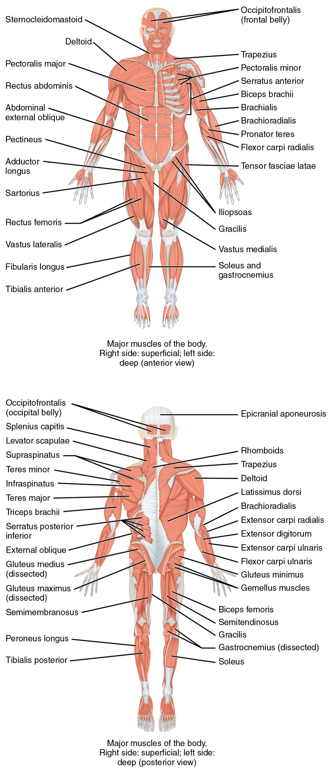
Figure 7.2 image description: The top panel shows the anterior view of the human body with the major muscles labeled. Labels read (from the top, head): occipitofrontalis (frontal belly), sternocleidomastoid, trapezius, deltoid, pectoralis minor, serratus anterior, pectoralis major, arm muscles: biceps brachii, brachialis, brachioradialis, pronator teres, flexor carpi radialis, abdominal: rectus abdominis, abdominal external oblique, lower body: tensor fasciae latae, iliopsoas, pectineus, adductor longus, sartorius, gracilis, rectus femoris, vastus lateralis, vastus medialis, fibularis longus, tibialis anterior. The bottom panel shows the posterior view of the human body with the major muscles labeled. Labels read (from the top, head, left side): epicranial aponeurosis, occipitofrontalis, splenius capitis, levator scapulae, rhombus, trapezius, supraspinatus, teres minor, infraspinatus, teres major, triceps brachii, serratus posterior inferior, external oblique, lower body: gluteus medius, gluteus maximus, semimembranosus, peroneus longus, tibialis posterior, (right side, from top) trapezius, deltoid, latissimus dorsi, arm: brachioradialis, extensor carpi radialis, extensor digitorum, extensor carpi ulnaris, flexor carpi ulnaris, lower body: gluteus minimus, gemellus muscles, biceps femoris, semitendinosus, gracilis, gastrocnemius, soleus. [Return to Figure 7.2].
Unless otherwise indicated, this chapter contains material adapted from Anatomy and Physiology (on OpenStax), by Betts et al. and is used under a CC BY 4.0 international license. Download and access this book for free at https://openstax.org/books/anatomy-and-physiology/pages/1-introduction.
definition
The muscles responsible for voluntary muscle movement; also called striated muscle (Betts et al., 2013)
Involuntary and found only in the heart. Highly coordinated contractions pump blood into the vessels of the circulatory system (Betts et al., 2013)
The muscles responsible for involuntary muscle movement; also called visceral or non-striated muscle (Betts et al., 2013)
The state of steady internal conditions maintained by living things (Betts et al., 2013)
In opposition to each other (Betts et al., 2013)
naming conventions
